

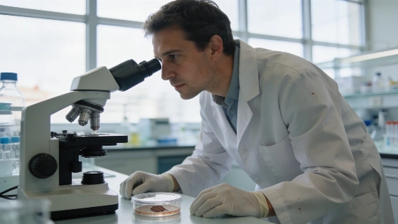
Researchers at Vall d’Hebron found that blocking p38α improved anti-PD-1 treatment in melanoma models. The work also identified a nine-gene signature linked to treatment response and survival.
In experiments led by the Vall d’Hebron Instituto de Investigación, melanoma tumours responded better to immunotherapy when researchers combined p38α inhibitors with anti-PD-1 treatment. Used alone, the inhibitors did not produce the same result. Some animal models showed complete tumour elimination.
The finding points to a treatment-resistance mechanism that combination therapy may help address.
Published in Nature Communications, the study examines how p38α affects melanoma growth and the immune environment around the tumour.
Researchers from VHIR’s Biomedical Research Group in Melanoma worked with specialists from Vall d’Hebron University Hospital’s Dermatology and Pathology departments. The VHIR group focused on childhood cancer and haematological diseases also took part, along with VHIO, Cemcat, CNAG and PRBB.
An earlier regional report covered a public event in Catalonia. This study examines a laboratory finding that may have a future role in cancer treatment.
p38α behaves differently at different stages of the disease. Before melanoma develops, the protein helps protect cells from damage caused by ultraviolet radiation. Once a tumour forms, p38α can support its growth and help create conditions that weaken the immune response.
The researchers removed p38α from tumour cells across animal models and cell cultures, and in human samples. CD8+ T lymphocytes and natural killer cells then became more active, while other signals linked to inflammation grew stronger.
The tumours became more sensitive to anti-PD-1 therapy, which is already used against melanoma. In the experiments, p38α inhibitors alone did not reduce tumour size. Combined with immunotherapy, they produced much better tumour control. Some animals reached complete tumour elimination.
VHIR researchers describe p38α as a possible target for overcoming resistance to immunotherapy. They say p38α inhibitors could make some less responsive tumours more vulnerable when used with anti-PD-1 treatment.
This is not yet a treatment option for patients. The findings need confirmation in more human samples, and further studies must assess the combination’s safety and effectiveness in clinical trials.
The study also points to a possible testing tool. Researchers identified a molecular signature of nine genes linked to p38α. Measuring the activity of those genes could help distinguish tumours more likely to respond to immunotherapy from those with a lower expected response.
The signature was also associated with better survival. For melanoma care, it suggests two possible uses: p38α could become a target for combination treatment, while the nine-gene profile could help with patient selection.
Until clinical testing is complete, the discovery remains an early research lead on immunotherapy resistance.