
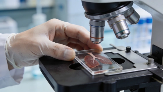
Researchers mapped 91,068 lung-tissue regions from 19 people who died with severe COVID-19. The analysis found stronger fibrosis-linked signals during a stage when alveolar repair was still underway.
The tissue samples came from 19 people who died with COVID-19. Researchers examined 91,068 microscopic regions and found that cell layout and communication changed as damage progressed. Seven patients had acute diffuse alveolar damage, or DAD, and 12 had the proliferative form. The analysis also included control lung tissue. In proliferative DAD, fibrosis-linked signals were stronger even as processes aimed at repairing the alveoli remained active.
The study involved the Institut de Recerca Sant Pau, or IR Sant Pau, in Barcelona and was published in Nature Communications. Manel Esteller led the work. He heads the cancer epigenetics group at IR Sant Pau and is an ICREA researcher. He is also a professor of genetics at the University of Barcelona. Carlos A. García-Prieto and Eva Musulén were the first authors. The Josep Carreras Leukaemia Research Institute took part, as did Barcelona Supercomputing Center. DAD is a form of lung injury associated with severe acute respiratory distress syndrome.
Spatial transcriptomics let the team track gene activity without losing its position in the tissue. Using the Human Lung Cell Atlas as a reference, they identified 45 cell types. Conventional transcriptomic analysis requires tissue to be broken apart and cells studied separately. This approach kept the location of each analysed region, so researchers could examine cell niches and signals exchanged by neighbouring cells across both DAD stages.
Alveolar type 1 cells became less common. They carry out much of the gas exchange between the lungs and blood.
In proliferative DAD, alveolar type 2 cells grew more abundant. They can multiply and act as progenitors for type 1 cells after injury. Their increase points to an active repair response against continuing tissue damage.
The researchers also found fewer capillary endothelial cells. Natural killer cells declined as well, while some macrophage populations increased. Peribronchial fibroblasts rose particularly sharply and formed distinct niches in the tissue. These cells produce components of the extracellular matrix. Their expansion was associated with the fibrotic response in advanced disease. As Manel Esteller told EFE, severe COVID-19 changes which cells are present, where they are distributed and what signals they exchange.
Cell-to-cell signalling analysis brought the TGF-β pathway into focus. It plays a role in tissue repair, but excessive or persistent activation can contribute to fibrosis. In proliferative DAD, SMAD3 activity increased. SMAD7, which restrains profibrotic TGF-β/SMAD3 signalling, decreased. TIMP1-CD63 also emerged as a prominent interaction in the profibrotic signature.
The team reconstructed a signalling network in peribronchial fibroblasts. The network linked activation of the ITGB1 receptor with changes in SMAD3 and SMAD7. It also showed activity in the kinase ILK, alongside AKT1. MAP2K4 was involved, as was MAPK14. Researchers say some of these components could be studied as potential targets for limiting lung fibrosis. The work analysed post-mortem tissue and does not show that blocking TGF-β, or any other pathway, improves patient outcomes.
Signs of attempted alveolar repair persisted even in advanced disease. By combining gene activity with cell position, the researchers identified different stages in the transition from type 2 to type 1 alveolar cells. Some regions contained cells at intermediate stages. Others showed CAV1, a marker of mature type 1 cells. Repair varied across the tissue and could remain incomplete.
Comparisons with eight cases of DAD unrelated to COVID-19 showed different fibroblast patterns. Peribronchial fibroblasts formed niches in proliferative COVID-19, while alveolar fibroblasts predominated in DAD from other causes. Peribronchial fibroblasts also appeared in idiopathic pulmonary fibrosis. In a separate comparison, data from more than 1.4 million Human Lung Cell Atlas cells showed that the SMAD3-high and SMAD7-low pattern was particularly associated with COVID-19.
The evidence has limits. Most samples came from people who had died, and the disease stages came from different patients, so the study cannot track progression within one person. Clinical differences between groups also make it difficult to separate infection-specific effects from comorbidities. Disease duration is another factor, as are secondary lung infections. The biological focus of this research is separate from the housing mobilisation report published earlier by RUSSPAIN.COM.